Study of cells: The prokaryotic cell, Archaea, Bacteria, The eukaryotic cell, Compartments
Study of cells
Biologists use various tools to achieve knowledge of the cells. They get information from their shapes, sizes and components, which allows them to further understand the functions performed within them. Since the first observations of cells for over 300 years until the present time, techniques and equipment have been refined, resulting in a branch of biology: the Microscopy. Given the small size of most cells, the use of the microscope is of great value in biological research. Today, biologists use two basic types of microscope: the optical and electronics.
The prokaryotic cell
Prokaryotic cells are small and less complex than eukaryotes. Contain ribosomes but lack endomembrane systems (ie, organelles delimited biological membranes, such as the cell nucleus). So have the genetic material in the cytosol. However, there are exceptions: some photosynthetic bacteria have internal membrane systems. Also in the Phylum Planctomycetes bodies exist as Pirellula surrounding its genetic material through a membrane and intracytoplasmic obscuriglobus Gemmata double surrounding membrane. The latter also has other internal membrane compartments, possibly connected with the outer membrane of the nucleoid and the nuclear membrane, which has no peptidoglycan.
Generally could be said to lack the prokaryotic cytoskeleton. However it has been observed that some bacteria such as Bacillus subtilis, possess proteins such as MreB and mbl acting in a manner similar to actin and are important in cell morphology. Fusinita van den Ent, in Nature, goes beyond stating that the cytoskeletons of actin and tubulin have prokaryotic origin.
Of great diversity, prokaryotes sustain metabolism extraordinarily complex, sometimes unique to certain taxa, as some groups of bacteria, which affects their versatility organic. The prokaryotes are classified according to Carl Woese, in archaea and bacteria .
Archaea
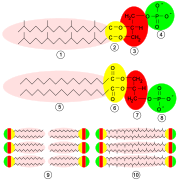
Structure biochemistry of archaea membrane (top) compared with bacteria and eukaryotes (middle): note the presence of ether linkages (2) replacing the ester (6) in phospholipids.
Archaea have a diameter between 0.1 and cell 15 ìm, although filamentous forms can be larger by aggregation of cells. Present a multitude of ways: including square and flat is described. Some archaea have flagella and are motile.
Archaea, like bacteria, have no internal membranes delimiting organelles. Like all organisms have ribosomes, but unlike those found in bacteria that are sensitive to certain antimicrobial agents, the archaeal, closer to eukaryotes, are not. The cell membrane has a structure similar to that of other cells, but its chemical composition is unique, with such ether bonds in lipids. Almost all archaea possess a cell wall (some Thermoplasma are the exception) ended property For example, do not contain peptidoglycan (murein), characteristic of bacteria. However, they may qualify under the stain of Gram, of vital importance in the taxonomy of bacteria, however, in archaea, possessing a wall structure common to all bacteria, the staining is applicable, but no taxonomic value. The order METHANOBACTERIALES has a layer of pseudomureína, which causes such as archaea respond positively to the Gram stain.
As in almost all prokaryotes, archaeal cells lack nuclei, and have a single circular chromosome. There extrachromosomal elements such as plasmids. Their genomes are small, about 2-4 million base pairs. Also characteristic is the presence of RNA polymerase complex and established a large number of nucleotides modified in the ribosomal ribonucleic acids. Moreover, its DNA is packaged in the form of nucleosomes, as in eukaryotes, because proteins similar to histones and some genes have introns. They can reproduce by binary fission or multiple fragmentation or budding.
Bacteria

Prokaryotic cell structure: 1, pili, 2, plasmid, 3, ribosomes, 4, cytoplasm; 5, plasma membrane; 6, cell wall; 7 capsule, 8, DNA, 9, bacterial flagellum.
Bacteria are relatively simple organisms, very small, barely a micron in most cases. Like other prokaryotes, lack a nucleus bounded by a membrane, but have a nucleoid, an elementary structure that usually contains a large circular molecule DNA. They have no nucleus and other organelles delimited by biological membranes. in the cytoplasm can be appreciated plasmids, small circular DNA molecules that coexist with the nucleoid and contain genes: they are commonly used by bacteria in the parasexual (sexual reproduction bacterial). The cytoplasm also contained ribosomes and various types of granules. In some cases there may be structures composed of membranes, usually related to photosynthesis.
They have a cell membrane composed of lipids, in the form of a bilayer and it is a cover on which there is a polysaccharide complex called peptidoglycan, depending on its structure and its response to subsequent Gram stain, are classified to the bacteria in Gram positive and Gram negative. The space between the cell membrane and cell wall (or outer membrane, if it exists) is called the periplasmic space. Some bacteria have a capsule. Others are able to produce endospores (dormant stages capable of withstanding extreme conditions) at some point in their life cycle. Among the external formations characteristic of the bacterial cell stand out scourges (of completely different structure of eukaryotic flagella) and pili (adhesion structures and related parasexual).
Most bacteria have a single circular chromosome and often have additional genetic elements, such as different types of plasmids. Reproduction, binary and very efficient in time, allows for rapid expansion of their populations, generating a large number of cells that are virtually clones, ie identical.
The eukaryotic cell
Eukaryotic cells are the exponent of the current cellular complexity. They have a relatively stable basic structure characterized by the presence of different types of organelles specialized intracytoplasmic, among which the nucleus, which houses the genetic material. Especially in multicellular organisms, cells can achieve a high degree of specialization. Such specialization or differentiation is such that, in some cases, compromises the viability of the cell type in isolation. Thus, for example, neurons depend for survival of glial cells.
Furthermore, the cell structure varies depending on the taxonomic status of the living: in this way, plant cells differ from animal as well as those of fungi. For example, animal cells lack a cell wall, are highly variable, not plastids, may have vacuoles but are not very large and have centrioles (which are aggregates of microtubules cylindrical contribute to the formation of cilia and flagella and facilitate the cell division). The cells of plants, meanwhile, have a cell wall made of cellulose), have plastids and chloroplasts (organelle capable of photosynthesis), chromoplasts (organelles that accumulate pigments) or Leucoplast (organelles that accumulate starch produced in photosynthesis), have vacuoles that accumulate large reserve substances or waste produced by the cell and eventually also have plasmodesmata, which are cytoplasmic connections that allow direct movement of substances in the cytoplasm of one cell to another, with continuity of their plasma membranes.
Compartments
Cells are dynamic entities, with cellular metabolism of highly active internal structure of which is a flow between routes anastomosed. A phenomenon observed in all cell types is compartmentalization, which consists of a heterogeneous environments resulting in more or less defined (surrounded or not by biological membranes) in which there is a microenvironment that brings together the elements involved in a biological pathway . This compartmentalization is demonstrated most clearly in eukaryotic cells, which consist of different structures and organelles that develop specific functions, which is a method of spatial and temporal specialization. However, simple cells, as prokaryotes, already have similar specializations.
Plasma membrane and cell surface
The plasma membrane composition varies depending on the function cells or tissue in which it is located, but has common elements. It consists of a double layer of phospholipids by proteins bound non-covalently to the bilayer, and carbohydrates attached covalently to lipids and proteins. Generally, larger molecules are the lipids, but the protein, due to its higher molecular mass, representing about 50% of the mass of the membrane.
A model that explains the operation of the plasma membrane is the fluid mosaic model of JS Singer and Garth Nicolson (1972), which develops a concept of unity based on thermodynamics hydrophobic interactions between molecules and other non-covalent bonds.
The membrane structure supports a complex mechanism of transport, which enables a fluid exchange of mass and energy between the intracellular and external environment. Moreover, the possibility of transport and interaction between neighboring cells or molecules in a cell with its environment enables them to communicate chemically, ie, allows cell signaling. neurotransmitters, hormones, local chemical mediators affecting specific cells by changing the pattern of gene expression through mechanisms of signal transduction.
On the lipid bilayer, irrespective of the presence or absence of a cell wall, there is a matrix that may vary from little conspicuous, as in the epithelium, very large, as in the connective tissue.

Like it on Facebook, Tweet it or share this article on other bookmarking websites.